Thursday 28 February 2013

Blood In Urine ladder Cancer

Source(google.com.pk)
Blood In Urine ladder Cancer
   Leukemia is a type of cancer which affects the bone marrow and is characterized by unrestrained proliferation of white blood cells. Its name “leukemia” means “white blood” since this cancer is characterized by a high number of white cells in the blood, many of which are immature or dysfunctional.

There are many types of leukemias, many of which develop as an acute (most frequent form in children) or a chronic disease.

Different leukemias have been linked to different causes, such as exposure to ionizing radiation or chemicals such as benzene or tobacco smoke. It can also develop as a consequence of chemotherapy for previous malignacies or viral infections (HTLV-1 virus). Some develop in people with chromosomal abnormalities or other genetic abnormalities.

Leukemia leads to the formation of metastatic colonies in other organs which requires a penetration of cancer cells through matrix barriers and blood vessel walls, a process mediated through MMPs

We investigated the effects of micronutrients on a number of human leukemia cells, including Adult T-cell leukemia (ATL), a fatal disease with an average survival time of less than one year, and  murine leukemia cells. In addition to using the micronutrient mixture we investigated the anti-cancer effects of some of its components individually, such as vitamin C, lysine and EGCG from green tea.

We have evaluated the effects of these micronutrients on limiting leukemia cancer cell growth, triggering the natural elimination of cancer cells by the induction of apoptosis (cell suicide), and decreasing cancer invasiveness by inhibition of  MMP secretion. In addition to the in vitro studies we also investigated the effects of micronutrient synergy on suppression of the tumor growth in nude mice.

Our results suggest that a specific combination of micronutrients containing vitamin C, Lysine, green tea extract rich in EGCG, and other natural components, is a promising new therapeutic agent for leukemia, and is a potential candidate for human trials.

Blood And Cancer

Source(google.com.pk)
Blood And Cancer
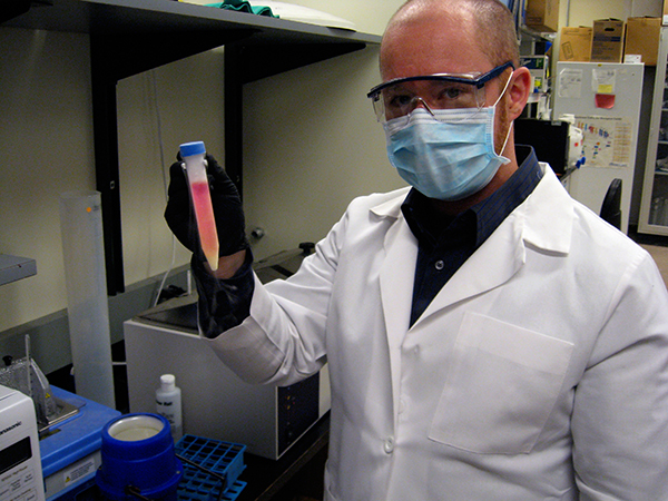
   In the battle against cancer, Dr. Judah Folkman (born 1933) has found a new approach: to attack the blood vessels that nourish cancer cells. The results of initial tests in cancer-bearing mice performed in 1998 were promising enough to raise the hopes of both cancer patients and physicians worldwide.
A noted professor, physician, and former surgeon-in-chief at the Boston Pediatric Hospital, Dr. Judah M. Folkman has demonstrated that by cutting off the blood supply nourishing cancer cells, cancer tumors can be killed with only negligible side effects. Theorizing on a method to prevent the growth of existing tumors, Folkman decided to try blocking the signals sent out by these tumors to perform angiogenesis - the formation of new blood vessels. To test his theory, he used two agents - angiostatin and endostatin - to treat cancer in laboratory mice. Although the results were positive, they were not conclusive, for other cancer-fighting drugs had a history of working well on mice but not as well on humans. By 2000 there remained the task of completing extensive tests on humans before Entre Med Inc., a Rockville, Maryland-based biotech company, could begin to put the new compounds on the market.

Discovered How Cancer Cells Grow
The son of Rabbi Jerome Folkman and his wife, Bessie, Folkman came to a love of medicine early in life. At his bar mitzvah his father told him to be a credit to his people; as an adult Folkman determined to dedicate himself to cancer research as a way of following his father's advice. In 1961 he made an astounding discovery. While doing medical research in a U.S. Navy laboratory, he found that cancer cells grow because they have an abundant blood supply. From this discovery Folkman developed the theory of angio-genesis and hypothesized further that cancers could not thrive without this abundant blood supply. A tumor formed, he theorized, because it could somehow stimulate new blood cells to deliver to it the nutrients it required in order to grow. Without sufficient blood, a cancer would not be able to grow any larger than a pin head.
Folkman's theory led him to look for ways to block blood vessel growth. He worked for years before he was able to develop compounds that sufficiently inhibited angiogenesis. These compounds, the human proteins endostatin and angiostatin, seemed like the answer to a prayer for people affected by cancer. Endostatin in particular showed promise; its marked lack of toxicity seemed to make it safe for human testing and two small-scale clinical trials at the U.S. National Cancer Institute (NCI) were authorized to further examine its effects on humans.
In February 1999, the NCI verified that Folkman's results with endostatin could be replicated. The human protein endostatin not only inhibited tumor growth in mice, but also showed no side effects when tested on in monkeys, even when administered in high doses. In an exciting announcement, the NCI confirmed that endostatin's anti-angiogenesis properties dramatically shrank cancerous tumors in laboratory mice.
The NCI's announcement came at the end of 30 years of arduous work on the part of Folkman. Years of work resulted in his discovery that blood vessels provided the key to cancer's survival, and much more research was subsequently undertaken in order for him to determine the basic process by which cancerous tumors spark the formation of the new blood vessels required to feed their growth. "Most research is failure," Folkman told NOVA producer Nancy Linde. "You go years and years and years, and then every once in a while there is a tremendous finding, and you realize for the first time in your life that you know something that hour or that day that nobody else in history has ever known, and you can understand something of how nature works."
Perhaps most frustrating for Folkman during his decades of research was the time it took him to convince the medical community that his theory had merit. Although he encountered skepticism from researchers, Folkman persisted because he knew that as a surgeon he had the hands-on experience with live human cancer tissues that researchers lacked. During surgery, Folkman had seen small tumors in the thyroid gland and the lining of the abdomen that never grew very large because they could not stimulate blood vessel growth. That led him to think that some kind of angiogenesis factor, possibly a diffusible protein, could stimulate the growth of blood vessels in certain tissues. But where the factor could not stimulate the formation of new blood vessels, the cancer did not grow. That meant cancer could be kept from growing if it could not attract the formation of a nutrient source. Folkman focused on determining what the factor was and if it could be blocked.
Discovered the Angiogenic Factor
Folkman maintains that being a researcher is one of the hardest jobs around, because, unlike a surgeon, a researcher doesn't get feedback from patients. That means that years of criticism, along with funding problems, must be faced before any positive reinforcement results from one's work. His own research experience was no exception to that rule. Fortunately, he was persistent; his knowledge as a practicing surgeon gave him the courage to continue presenting his ideas.
Although Folkman's ideas were at first largely discounted, by the 1970s that began to change. Researchers agreed that tumors did seem to cause the presence of new blood vessels, but most thought it was a side effect of dying cancer cells. They did not believe that the live cells actually stimulated the formation of new blood vessels.
When Robert Auerbach came to Folkman's lab, the two researchers conducted an experiment that proved the cancer cells were in fact causing the formation of blood vessels as a means of feeding their growth. Auerbach put live tumor cells in one eye of a rabbit and dying tumor cells in the other eye. The results showed that blood vessels formed around the live cells, not the dying ones. This proved that live cancer cells were actually causing the growth of the blood vessels.
In 1971 the New England Journal of Medicine published Folkman's paper that discussed the angiogenesis factor. Folkman began by noting that new blood vessels were recruited by tumors. Second, he maintained, the tumors sent out a factor that caused angiogenesis, or the formation of new blood vessels. Third, he said, this factor would stimulate the growth of new blood vessels. If this factor could be blocked, he hypothesized, tumors would stay small.
In 1984 Folkman and his team of researchers published a paper about the first angiogenic factor, a molecule that stimulated angiogenesis. He was later to discover 17 molecules, but the discovery of the first convinced him that he was on the right track. When he was unable to find the biological factor that stimulated blood vessel growth, Folkman began to wonder if he was being headstrong about his ideas rather than persistent. Realizing that he was right took time, but it was worth it in the end, when the medical community realized that through his work there was new hope for cancer patients: their disease could be stopped without debilitating side effects.
Experimented with Inhibitors
Killing cancer is a daunting task that often requires extensive drug treatments, chemotherapy, and radiation, which takes a heavy toll on healthy cells in the body. Cancer cells are more resistant to drugs than are normal cells, which is part of the reason why they are so hard to kill. The genius of Folkman's solution is that blood vessels are normal cells that respond readily to drugs. When subjected to angiostatin and endostatin treatments, blood vessels disappear, and with them, the tumors that feed on them. The anti-angiogenesis process might be compared to starving an enemy out by laying siege to his castle. No food supplies can get in, so the enemy eventually weakens. "We have been leveling out in our ability to stop cancer using available tools," Robert Siegel, director of hematology and oncology at George Washington University Medical Center, told Insight on the News: "The idea of having a completely new approach that is effective with some cancers is exciting."
Following testing on endostatin and its approval for use on human subjects, Duane Gay was one of the first to be treated with the substance. Although a tumor in his rib grew, those in his liver, lungs, and kidney stabilized. With the anti-angiogenic benefits of the drug interferon-alpha, Folkman also had successes treating children with hemangiomas, or life-threatening masses of blood capillaries. By the year 2000, 30 drugs were considered angiogenic inhibitors.
The Value of Observation
That Folkman perceived something no one else had noticed while working with cancer patients was not surprising to the people who know him best. Dr. C. Everett Koop, one of Folkman's colleagues at Boston Children's Hospital, found his colleague's power of observation startling. For example, when Folkman started in pediatric surgery, he had no experience working on young children. Koop painstakingly showed him the difference between pediatric tissues and adult tissues. While watching Koop, Folkman discovered that Koop's technique largely depended on steadying things with his left thumbnail against his left forefinger. Fingernails, Folkman decided, made all the difference. Koop had never realized how important his fingernails were before, but he knew Folkman was right.
Folkman's skill as a pediatric surgeon grew, as did his skill as a physician and researcher. He also becames a noteworthy professor whose students at Harvard Medical School counted him among their best teachers. Throughout the years Folkman conveyed to his students the importance of staying connected to patients. As a doctor, he gave his home telephone number and beeper number so that his patients could contact him. As a surgeon, he believed there was no such thing as "false hope." He advised his students never to tell a patient that there was nothing they could do to help him or her, because there was always something, even if it was only making the patient feel better.
In all his years of research, Folkman never lost sight of the people he was helping. Despite repeated criticism, he persevered with his assertions regarding cancer treatments until the medical community finally acknowledged that his research had in fact expanded their understanding of the disease. In fact, Folkman's research has applications for 26 diseases, including arthritis, cancer, Crohn's disease, endometriosis, and leukemia.
Robert Cooke, author of Dr. Folkman's War: Angiogenesis and the Struggle to Defeat Cancer, wrote that by 2000 Folkman's work was clearly recognized as having contributed to the sum of medical knowledge. "Finally it seemed that his peers were judging him to be persistent, not obstinate," Cooke wrote. "This was a distinction he had long sought. Now it seemed clear that great strides had been made largely because one man worked, pushed, and badgered one idea for so many years. Step by painful step, at first alone and then with colleagues he had engaged the struggle, Folkman had faced the objections and surmounted all the barriers that inflexible critics and doubters threw in his path. This experience had bred an enduring confidence and had even given him a sense of peace."
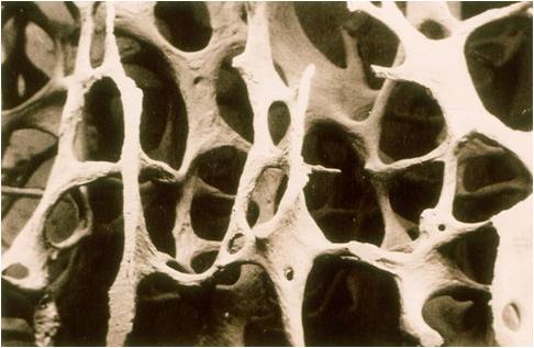
Acute Leukemia

Source(google.com.pk)
Acute Leukemia

Leukemia (British spelling: leukaemia ) is cancer of the blood or bone marrow (which produces blood cells). A person who has leukemia suffers from an abnormal production of blood cells, generally leukocytes (white blood cells).
The word Leukemia comes from the Greek leukos which means "white" and aima which means "blood".

The DNA of immature blood cells, mainly white cells, becomes damaged in some way. This abnormality causes the blood cells to grow and divide chaotically. Normal blood cells die after a while and are replaced by new cells which are produced in the bone marrow. The abnormal blood cells do not die so easily, and accumulate, occupying more and more space. As more and more space is occupied by these faulty blood cells there is less and less space for the normal cells - and the sufferer becomes ill. Quite simply, the bad cells crowd out the good cells in the blood.

In order to better understand what goes on we need to have a look at what the bone marrow does.

Function of the bone marrow
The bone marrow is found in the inside of bones. The marrow in the large bones of adults produces blood cells. Approximately 4% of our total bodyweight consists of bone marrow.

There are two types of bone marrow: 1. Red marrow, made up mainly of myeloid tissue. 2. Yellow marrow, made up mostly of fat cells. Red marrow can be found in the flat bones, such as the breast bone, skull, vertebrae, shoulder blades, hip bone and ribs. Red marrow can also be found at the ends of long bones, such as the humerus and femur.

White blood cells (lymphocytes), red blood cells and platelets are produced in the red marrow. Red blood cells carry oxygen, white blood cells fight diseases. Platelets are essential for blood clotting. Yellow marrow can be found in the inside of the middle section of long bones.

If a person loses a lot of blood the body can convert yellow marrow to red marrow in order to raise blood cell production.

White blood cells, red blood cells and platelets exist in plasma - Blood plasma is the liquid component of blood, in which the blood cells are suspended.
Types of leukemia

Chronic and Acute
Experts divide leukemia into four large groups, each of which can be Acute, which is a rapidly progressing disease that results in the accumulation of immature, useless cells in the marrow and blood, or Chronic, which progresses more slowly and allows more mature, useful cells to be made. In other words, acute leukemia crowds out the good cells more quickly than chronic leukemia.

Lymphocytic and Myelogenous
Leukemias are also subdivided into the type of affected blood cell. If the cancerous transformation occurs in the type of marrow that makes lymphocytes, the disease is called lymphocytic leukemia. A lymphocyte is a kind of white blood cell inside your vertebrae immune system. If the cancerous change occurs in the type of marrow cells that go on to produce red blood cells, other types of white cells, and platelets, the disease is called myelogenous leukemia.

Acute Lymphocytic Leukemia (ALL), also known as Acute Lymphoblastic Leukemia - This is the most common type of leukemia among young children, although adults can get it as well, especially those over the age of 65. Survival rates of at least five years range from 85% among children and 50% among adults. The following are all subtypes of this leukemia: precursor B acute lymphoblastic leukemia, precursor T acute lymphoblastic leukemia, Burkitt's leukemia, and acute biphenotypic leukemia.

Chronic Lymphocytic Leukemia (CLL) - This is most common among adults over 55, although younger adults can get it as well. CLL hardly ever affects children. The majority of patients with CLL are men, over 60%. 75% of treated CLL patients survive for over five years. Experts say CLL is incurable. A more aggressive form of CLL is B-cell prolymphocytic leukemia.

Acute Myelogenous Leukemia (AML) - AML is more common among adults than children, and affects males significantly more often than females. Patients are treated with chemotherapy. 40% of treated patients survive for over 5 years. The following are subtypes of AMS - acute promyelocytic leukemia, acute myeloblastic leukemia, and acute megakaryoblastic leukemia.

Researchers from the Memorial Sloan-Kettering Cancer Center reported in the March 2012 issue of NEJM (New England Journal of Medicine that they identified a series of genetic mutations in people with AML. They explained that their findings may help doctors to more accurately predict patient outcomes, as well as choosing therapies they are most likely to respond to.

Chronic Myelogenous Leukemia (CML) - The vast majority of patients are adults. 90% of treated patients survive for over 5 years. Gleevec (imatinib) is commonly used to treat CML, as well as some other drugs. Chronic monocytic leukemia is a subtype of CML.

Symptoms of leukemia
Blood clotting is poor - As immature white blood cells crowd out blood platelets, which are crucial for blood clotting, the patient may bruise or bleed easily and heal slowly - he may also develop petechiae (a small red to purple spot on the body, caused by a minor hemorrhage).

Affected immune system - The patient's white blood cells, which are crucial for fighting off infection, may be suppressed or not working properly. The patient may experience frequent infections, or his immune system may attack other good body cells.

Anemia - As the shortage of good red blood cells grows the patient may suffer from anemia - this may lead to difficult or labored respiration (dyspnea) and pallor (skin has a pale color caused by illness).

Other symptoms - Patients may also experience nausea, fever, chills, night sweats, flu-like symptoms, and tiredness. If the liver or spleen becomes enlarged the patient may feel full and will eat less, resulting in weight loss. Headache is more common among patients whose cancerous cells have invaded the CNS (central nervous system).

Precaution - As all these symptoms could be due to other illnesses. A diagnosis of leukemia can only be confirmed after medical tests are carried out.

What causes leukemia?
Experts say that different leukemias have different causes. The following are either known causes, or strongly suspected causes:
Artificial ionizing radiation
Viruses - HTLV-1 (human T-lymphotropic virus) and HIV (human immunodeficiency virus)
Benzene and some petrochemicals
Alkylating chemotherapy agents used in previous cancers
Maternal fetal transmission (rare)
Hair dyes
Genetic predisposition - some studies researching family history and looking at twins have indicated that some people have a higher risk of developing leukemia because of a single gene or multiple genes.
Down syndrome - people with Down syndrome have a significantly higher risk of developing leukemia, compared to people who do not have Down syndrome. Experts say that because of this, people with certain chromosomal abnormalities may have a higher risk.
Electromagnetic energy - studies indicate there is not enough evidence to show that ELF magnetic (not electric) fields that exist currently might cause leukemia. The IARC (International Agency for Research on Cancer) says that studies which indicate there is a risk tend to be biased and unreliable.

Treatments for leukemia
As the various types of leukemias affect patients differently, their treatments depend on what type of leukemia they have. The type of treatment will also depend on the patient's age and his state of health.

In order to get the most effective treatment the patient should get treatment at a center where doctors have experience and are well trained in treating leukemia patients. As treatment has improved, the aim of virtually all health care professionals should be complete remission - that the cancer goes away completely for a minimum of five years after treatment.

Treatment for patients with acute leukemias should start as soon as possible - this usually involves induction therapy with chemotherapy, and takes place in a hospital.

When a patient is in remission he will still need consolidation therapy or post induction therapy. This may involve chemotherapy, as well as a bone marrow transplant (allogeneic stem cell transplantation).

If a patient has Chronic Myelogenous Leukemia (CML) his treatment should start as soon as the diagnosis is confirmed. He will be given a drug, probably Gleevec (imatinib mesylate), which blocks the BCR-ABL cancer gene. Gleevec stops the CML from getting worse, but does not cure it. There are other drugs, such as Sprycel (dasatinib) and Tarigna (nilotinb), which also block the BCR-ABL cancer gene. Patients who have not had success with Gleevec are usually given Sprycel and Tarigna. All three drugs are taken orally. A bone marrow transplant is the only current way of curing a patient with CML. The younger the patient is the more likely the transplant will be successful.

Synribo (omacetaxine mepesuccinate) was approved by the FDA, on 26th October 2012, for the treatment of chronic myelogenous leukemia (CML) in adult patients who had been treated with at least two drugs, but whose cancer continued to progress. Resistance to medications is common in CML. Synribo is an alkaloid from Cephalotaxus harringtonia which inhibits proteins that trigger the development of cancerous cells. The drug is administered subcutaneously.

Patients with Chronic Lymphocytic Leukemia (CLL) may not receive any treatment for a long time after diagnosis. Those who do will normally be given chemotherapy or monoclonal antibody therapy. Some patients with CLL may benefit from allogeneic stem cell transplantation (bone marrow transplant).

Rabbit antibodies help Leukemia patients - scientists from Virginia Commonwealth University reported in the journal Bone Marrow Transplantation (July 2012 issue) that rabbit antibodies can improve survival and reduce the occurrence of relapses in patients with leukemia and myelodysplasia who are receiving a stem transplant from an unrelated donor.

Leukemia patients' own T-cells achieve remission for over two years - patients who were infused with their own T-cells after they had been genetically altered to fight cancer tumors stayed in full remission for over 24 months. Researchers from the Perelman School of Medicine at the University of Pennsylvania presented their findings at the American Society of Hematology's Annual Meeting and Exposition in December 2012. All those who took part in the human study had advanced cancers - ten of them had chronic lymphocytic leukemia, and two children had acute lymphoblastic leukemia.

All leukemia patients, regardless of what type they have or had, will need to be checked regularly by their doctors after the cancer has gone (in remission). They will undergo exams and blood tests. The doctors will occasionally test their bone marrow. As time passes and the patient continues to remain free of leukemia the doctor may decide to lengthen the intervals between tests.

About Leukemia

Source(google.com.pk)
About Leukemia

Leukemia is a cancer of the bone marrow and blood. There are four main types of leukemia. These are: acute lymphocytic leukemia (ALL), chronic lymphocytic leukemia (CLL), acute myelogenous leukemia (AML), and chronic myelogenous leukemia (CML). MLL stands for “mixed lineage leukemia” and means that the leukemia comes from both the myeloid and the lymphoid cell progenitors (Robien and Ulrich, 2003). The cause of leukemia is currently still unknown. It often arises as a result of DNA translocations, inversions, or deletions in genes regulating blood cell development or homeostasis. In all types of leukemia, genetic translocations, inversions, or deletions cause dysfunctional cells to replace normal hematopoietic cells in the bone marrow (Robien and Ulrich, 2003). A leukemia patient will usually die from anemia or infection because of the lack of red blood cells and immune cells (Sompayrac, 1999).

Leukemia stem cells (LSC) are thought to have been derived from haematopoietic stem cells (HSC), which are CD34+CD38-. During leukemogenesis, the LSC expresses shared surface characteristics with the HSC. It also has the capacity for producing both the clonogenic leukemic progenitors and the non-clonogenic blast cells, which make up the bulk of the leukemia (Huntly, et al. 2005). In the bone marrow there are two types of hematopoietic stem cells. In someone who does not have leukemia, the myeloid progenitor is the parent cell to granulocytes and macrophages, and the lymphoid progenitor is the parent cell to T-cells and B-cells (Janeway, et al., 1999). In adults, 85% of all leukemia cases are myeloid, with 15% being lymphoid. In children the opposite is true, with 80% of all cases being lymphoid (ALL) and only 20% being myeloid (Greaves, 2000).

In people with acute leukemia, a mistake is made in the action of the VDJ recombinase enzyme, which normally creates antibody and T-cell receptor diversity. Proto-oncogene, a gene that promotes growth and spread, is inappropriately turned on and it activates other proto-oncogenes and deactivate anti-oncogenes, which normally protect cells against cancer-causing mutations (Sompayrac, 1999). Chronic leukemias may be caused when mistakes in recombination activate genes that either cause the cell to proliferate, or cause it not to die by apoptosis or increased activity of stem cells or abnormal committed progenitor cells (Marley and Gordon, 2005). The increased life span of the cell increases the chance that enough mutations will accumulate to cause cancer (Sompayrac, 1999).

Mutations that increase the risk of leukemia can be caused by radiation, extremely low frequency electromagnetic fields, pesticides, benzene, other carcinogens, viral infections, and recombination errors that occur throughout life (Sompayrac, 1999). Cigarette smoke, high altitude, or other factors that increase exposure to radiation can be accelerating factors for cancer (Sompayrac, 1999). Rates of leukemia may also be greater following periods of population mixing. During periods of population mixing, individuals are exposed to new infectious agents. A similar hypothesis is that children who were not exposed to common infectious agents at a young age would also have higher rates of leukemia. The infectious agents would be new to their system, causing a strong and inappropriate immune response that might trigger leukemia. Studies on daycare attendance as a measure of exposure to infectious agents have had mixed results (Robien and Ulrich, 2003).

Immuno-suppressed people such as patients being treated with chemotherapy or patients who have AIDS have higher rates of leukemia, but they do not have higher rates of other types of cancer (Sompayrac, 1999). Some cells in the immune system may protect against the development of cancer. Macrophages are cells that eat and destroy old or damaged red blood cells. The macrophages recognize the cells because a fat molecule called phospotidyl-serine flips to the outside of the cell as it ages (Sompayrac, 1999). Macrophages can also destroy cancer cells, but only when they are hyperactivated. Usually when there is no inflammatory reaction in the body, the macrophages will remain resting and will not attack the cancer cells, but natural killer cells secrete cytokines when cancer cells are present and the cytokines cause the hyperactivation of the macrophages (Sompayrac, 1999). The immune system has evolved to protect against leukemia by destroying cancerous cells, but in a person whose immune system is weakened or is being exposed to many new pathogens, it is not as effective at getting rid of damaged cells.

Specific Types of Leukemia
ALL is a disease of B or T lymphocyte lineage. Childhood ALL is of the B lineage. One of the most common translocations in B-precursor childhood ALL is suggested to be t(1;19)(q23;p13). The deletion of 9p has been suggested to be an evolutionary aspect of the progression of ALL but it is also thought to play a primary role in some cases of leukemogenesis (Forestier, et al., 2000). In ALL, it seems likely that leukemia develops in two stages: a pre-natal genetic alteration that predisposes the infant to developing leukemic precursor cells, and a post-natal event that triggers this latent disease. This has been suggested by several studies, including a number of "twin studies," which compare the incidence of diseases in identical (monozygotic) and fraternal (dizygotic) twins, in order to determine the relative importance of genetics and the environment as causal factors in a specific disease. Many twin studies have shown that identical twins have high "concordance" levels for leukemia, meaning that they share the disease. Concordance has often been taken as proof that a disease is genetic. However, in this case, it may be because they share a very similar environment during their fetal development: more than half of identical twins share a placenta, while no fraternal twins do (Greaves, 1999). The leukemic genetic alteration most likely takes place in one twin and spreads to the other through the placenta. This suggests that in non-twin children, the leukemic alteration may also take place in utero, as indicated by positive results from blood samples taken soon after birth (Greaves, 1999). Concordance rates near 100% would imply that the initial leukemic genetic alteration is the sole and sufficient cause for the development of leukemia. This is the case with another type of leukemia, MLL. However, in ALL, the concordance rate is closer to 10%. This suggests that the initial genetic alteration is not enough to cause leukemia in the absence of a second genetic alteration caused by the post-natal environment (Greaves, et al, 2003).

CML is caused by the reproduction of cells that have not matured properly, but continue to reproduce (Marley and Gordon, 2005). It has a distinguishing inherited characteristic called the Philadelphia chromosome (No. 22), a genetic abnormality in the blood cells that is referred to as the Ph-chromosome.  The breakage on the chromosome is referred to as "BCR" (breakpoint cluster region). A breakage on chromosome 9, known as "ABL" (Abelson) has also been noted. These two mutated genes fuse together, forming a gene called BCR-ABL.  This gene can still function properly. However, in CML patients, the protein that is produced is abnormal, causing unregulated myeloid cell production. Evidence has pointed to this abnormal protein production as the cause of the leukemic conversion of the hematopoietic stem cell (Leukemia and Lymphoma Society, 2005). Normally stem cells divide in the bone marrow to replace themselves and create differentiating cells. In CML, either the stem cells or progenitor cells are increasing at all times (Marley and Gordon, 2005).

The most common genetic abnormality in CLL is 13q14 deletion, observed in 50% of all cases. (Caporaso et al., 2004)

In addition to the four basic types of leukemia, there are a variety of other forms. Different types of leukemia are characterized by different patterns of nonrandom chromosomal aberrations, but the frequencies with which the various karyotypic subtypes are seen differ amongst geographic regions. In areas where children are not sufficiently exposed in early youth to childhood infections, there is a greater risk of developing leukemia from an abnormal immune system response.

A Lymphoma

Source(google.com.pk)
A Lymphoma
  I started a new treatment in March, a week late, as I had to help move my grandma, since only one of her 6 children (plus 5 spouses) would help. My bf was going to help, but she changed the moving date, and he had already booked a day off for moving her.
So my mother and I were the only ones there to move her.

In April, I woke up with a swollen face, for no particular reason. Or at least, no reason was found.

I got a hickman line for my treatment, because my veins are so small, etc. I feel sorry for the nurses who have to find my veins. I have freaked a few out, causing them to poke and search numerous times for ONE IV insertion.

In May, the line got infected… I took a road trip to Wichita, Kansas from Buffalo, NY. 20 hour drive! With the stops for food, gas, rest, etc. it took 24 hours. I was very excited that my time estimation was right on! And I over estimated the fuel costs, so we had a good trip overall.

I woke up again beginning of June, with a swollen face. This time it was more intense, and went down to my throat. It was so bad that it altered my voice! I went to the local urgent care, and got a shot of Benedryl and a steroid shot. One in each buttock! OUCH!
I ended up in hospital for a week in June, for an undetermined infection. That was “fun”! It was during the 2nd week of my daughter’s summer visit. But I called her everyday, a few times a day. My friend was also going through a rough time, due to domestic abuse.

My hickman line was removed in July. That took a while to heal, due to the infection.

August was pretty uneventful, thankfully. August was also the end of my treatment.

First weekend of September, I spiked a fever of 105! Again I was admitted to hospital for a week. The doctor I had that time was insane, inconsiderate and useless. She did some of the stupidest things. Didn’t give me blood until 3 days after being there, when I went in with a low hemoglobin, and it dropped further the next day. Then she gave me something after the blood to help flush the excess fluids… this was at MIDNIGHT! The next day I had to take medical transport 2 hours away to go see my oncologist, so due to the medication she gave me to flush the fluids, we had to stop, so we were late. Didn’t help that the transport arrived to pick me up late, AND we had to stop for GAS! Crazy people!

October was somewhat crazy, because I was moving, and no one showed up to help, so it took my bf and I 2 weeks to move our stuff across the street. We were supposed to have the last 2 weeks of September to move our things into the new place, but the maintenance guy didn’t finish things until the last week, and left such a huge mess, that it took me 2 days to clean up after him! OH! To add to the stress of moving, I’d had a spot on my leg for over a month, that initially I thought was a mosquito bite, but it wasn’t itchy, so I eventually forgot about it. Well, suddenly, one day at the beginning of October, it started to itch, then a day later, it got sore. The next day it started to swell! I went to the urgent care again, for this. The doctor’s assumption was an ingrown hair. She told me that she could not be sure, without cutting it open. But she gave me antibiotics (At this point, I’d pretty much been on some form of antibiotics for the past 4-6 months!). A couple days later, it had swollen to the size of a quarter, and was at least half an inch high. It could barely walk (oh yeah, it was just to the front of my inner thigh). Finally, I’d had enough. I burst it open, because the pressure was SO intense! Oh my goodness, the nasty puss that came out of that thing, and the amount of it. WOW! Immediate relief from the pressure. But now I had to keep it clean, by flushing with saline solution. Thankfully, I had some left from when I’d had the hickman line. There is still a mark, I don’t think it will ever go away.

November was ok. December was my birthday and Christmas. And the year is done. We are not doing any testing beyond blood work, unless/until my symptoms return.

OH, and after all the years of chemotherapy, radiation, stem cell transplant, etc. I am finally gone into menopause. These hot flashes and mood swings are awful, and I feel very sorry for my wonderful boyfriend. Thankfully, he is wonderful; he has been amazing through all this stuff. I appreciate him more than I could ever express.

Cancer Bone Health

Source(google.com.pk)
Cancer Bone Health
Cancers can be discovered in bones in a number of different circumstances. When cancer is located in the bones, it is important to differentiate whether this cancer has spread from another site to the bones or whether the cancer originated in the bone tissue itself. This distinction is important not only for the sake of correct terminology, but also to accurately determine which treatment options are appropriate.
There are more than 100 types of cancer, and each cancer type is named for the organ or tissue in which it begins. When cancer cells spread, they may travel via the lymphatic channels to lymph nodes, or they may enter the bloodstream and travel to other organs or locations in the body that are distant from the site of the original, or primary, tumor. It is not unusual for cancers that originate in other parts of the body to spread to the bones and begin growing there. Certain types of cancers are particularly likely to spread to the bones. Cancers that commonly metastasize, or spread, to the bones include breast cancer, lung cancer, thyroid cancer, prostate cancer, and cancers of the kidney.

It is important to note that when these other types of cancer spread to the bone, they are still named for the tissue or organ where they arose and are not termed "bone" cancer. For example, breast cancer that has spread to the bones is referred to as metastatic breast cancer and not bone cancer.

In contrast to cancers which have spread to the bone, true bone cancers are tumors that arise from the tissues of the bones. These cancers, called primary bone cancers, are quite rare in comparison to cancers that have spread to the bones.

True bone cancer affects over 2,000 people in the United States each year. It is found most often in the bones of the arms and legs, but it can occur in any bone. Children and young people are more likely than adults to develop bone cancer. The symptoms of bone cancer tend to develop slowly and depend on the type, location, and size of the tumor. Pain is the most frequent symptom of bone cancer, but sometimes a lump on the bone can be felt through the skin.

Bone cancers are known as sarcomas. There are several types of sarcomas of bone, depending upon the kind of bone tissue where the tumor developed. The most common types of primary bone cancer are osteosarcoma, Ewing's sarcoma, and chondrosarcoma.

Osteosarcoma usually occurs between ages 10 and 25 and is more common in males than in females. Osteoosarcoma is most commonly found in the long bones of the arms or legs. Ewing's sarcoma is a tumor most commonly seen in people between 10 and 25 years of age that develops in the shaft (middle portion) of large bones, such as the hip bones, the long bones of the legs and upper arms, and the ribs. Chondrosarcoma is the bone tumor that is found mainly in adults. Chondrosarcoma arises from cartilage, the tissue located around the joints. Other, more rare types of bone cancer include fibrosarcoma, malignant giant cell tumor, and chordoma.

The treatment and prognosis of bone cancer depend upon multiple factors including the type and extent of the cancer, the patient's age and overall health status. Bone cancer may be treated with surgery, radiation therapy, chemotherapy, or a combination of these. Treatment for cancer that has spread to the bones (metastatic cancer) depends on the type of cancer (the tissue where the cancer started) and the extent of the spread.

Finally, another type of cancer can arise from blood cells produced in the bone marrow. Leukemia, multiple myeloma, and lymphoma are cancers that arise from these cells. These are referred to as hematologic malignancies (cancers of blood cells) and are not types of bone cancer.
Cancer Bone Health
Cancer Bone Health
Cancer Bone Health
Cancer Bone Health
Cancer Bone Health
Cancer Bone Health
Cancer Bone Health
Cancer Bone Health
Cancer Bone Health
Cancer Bone Health
Cancer Bone Health

Wednesday 20 February 2013

Daily Health Tips

Source(google.com.pk)
Daily Health Tips
Experts agree the key to healthy eating is the time-tested advice of balance, variety and moderation. In short, that means eating a wide variety of foods without getting too many calories or too much of any one nutrient. These 10 tips can help you follow that advice while still enjoying the foods you eat.


See Reference Guide for: Vitamins   Minerals   Amino Acids   Herbs   Special Nutrients   Anti-Oxidants  
 Dietary Fibers   Nutritional Greens   Digestive Nutrients   also see   Nutritional Glossary   INDEX 

Eat a variety of nutrient-rich foods. You need more than 40 different nutrients for good health, and no single food supplies them all. Your daily food selection should include bread and other whole-grain products; fruits; vegetables; dairy products; and meat, poultry, fish and other protein foods. How much you should eat depends on your calorie needs. Use the Food Guide Pyramid and the Nutrition Facts panel on food labels as handy references.
Enjoy plenty of whole grains, fruits and vegetables. Surveys show most Americans don't eat enough of these foods. Do you eat 6-11 servings from the bread, rice, cereal and pasta group, 3 of which should be whole grains? Do you eat 2-4 servings of fruit and 3-5 servings of vegetables? If you don't enjoy some of these at first, give them another chance. Look through cookbooks for tasty ways to prepare unfamiliar foods.
Maintain a healthy weight. The weight that's right for you depends on many factors including your sex, height, age and heredity. Excess body fat increases your chances for high blood pressure, heart disease, stroke, diabetes, some types of cancer and other illnesses. But being too thin can increase your risk for osteoporosis, menstrual irregularities and other health problems. If you're constantly losing and regaining weight, a registered dietitian can help you develop sensible eating habits for successful weight management. Regular exercise is also important to maintaining a healthy weight.
Eat moderate portions. If you keep portion sizes reasonable, it's easier to eat the foods you want and stay healthy. Did you know the recommended serving of cooked meat is 3 ounces, similar in size to a deck of playing cards? A medium piece of fruit is 1 serving and a cup of pasta equals 2 servings. A pint of ice cream contains 4 servings. Refer to the Food Guide Pyramid for information on recommended serving sizes.
Eat regular meals. Skipping meals can lead to out-of-control hunger, often resulting in overeating. When you're very hungry, it's also tempting to forget about good nutrition. Snacking between meals can help curb hunger, but don't eat so much that your snack becomes an entire meal.
Reduce, don't eliminate certain foods. Most people eat for pleasure as well as nutrition. If your favorite foods are high in fat, salt or sugar, the key is moderating how much of these foods you eat and how often you eat them.
Identify major sources of these ingredients in your diet and make changes, if necessary. Adults who eat high-fat meats or whole-milk dairy products at every meal are probably eating too much fat. Use the Nutrition Facts panel on the food label to help balance your choices.
Choosing skim or low-fat dairy products and lean cuts of meat such as flank steak and beef round can reduce fat intake significantly.
If you love fried chicken, however, you don't have to give it up. Just eat it less often. When dining out, share it with a friend, ask for a take-home bag or a smaller portion.
Balance your food choices over time. Not every food has to be "perfect." When eating a food high in fat, salt or sugar, select other foods that are low in these ingredients. If you miss out on any food group one day, make up for it the next. Your food choices over several days should fit together into a healthy pattern.
Know your diet pitfalls. To improve your eating habits, you first have to know what's wrong with them. Write down everything you eat for three days. Then check your list according to the rest of these tips. Do you add a lot of butter, creamy sauces or salad dressings? Rather than eliminating these foods, just cut back your portions. Are you getting enough fruits and vegetables? If not, you may be missing out on vital nutrients.
Make changes gradually. Just as there are no "superfoods" or easy answers to a healthy diet, don't expect to totally revamp your eating habits overnight. Changing too much, too fast can get in the way of success. Begin to remedy excesses or deficiencies with modest changes that can add up to positive, lifelong eating habits. For instance, if you don't like the taste of skim milk, try low-fat. Eventually you may find you like skim, too.
Remember, foods are not good or bad. Select foods based on your total eating patterns, not whether any individual food is "good" or "bad." Don't feel guilty if you love foods such as apple pie, potato chips, candy bars or ice cream. Eat them in moderation, and choose other foods to provide the balance and variety that are vital to good health.

Daily Health Tips
Daily Health Tips
Daily Health Tips
Daily Health Tips
Daily Health Tips
Daily Health Tips
Daily Health Tips
Daily Health Tips
Daily Health Tips
Daily Health Tips
Daily Health Tips
Daily Health Tips

Friday 15 February 2013

About Brain Cancer

Source(google.com.pk)
About Brain Cancer
Brain tumors are not rare.  Each year more than a 100,000 Americans will be diagnosed with a primary brain tumor or metastatic (secondary) brain tumor. Being diagnosed with a brain tumor can cause shock, fear, and confusion. For most people, having a brain tumor raises a lot of questions about what to do, where to go, and how to find the most comprehensive care. Dr. Sperduto and his expert team are here to help. From diagnosis to treatment, our experienced, concerned team of experts specializes in the comprehensive care of brain tumors.
The brain is the most important organ in the body.  It controls the five senses as well as the ability to speak and move.  In this section you will find a brief introduction to: brain anatomy,brain tumors, the diagnosis of brain tumors, and a description of the different types of brain tumors.
-------------------------------------------------------------------
BRAIN ANATOMY 
Central Nervous System (CNS): Your CNS consists of the brain and spinal cord.
Brain Stem: Connects your brain with your spinal cord and controls your breathing and heart rate.
Cerebral Cortex: Involved in your thinking, learning and speaking activities.

Cerebellum: Like a mini-brain within your brain, your cerebellum allows you to carry out skilled, complicated movements, including balance. 

Cerebrospinal fluid: Protects your brain and spinal cord by acting as a shock absorber.

Cerebrum: The largest and uppermost portion of your brain. It consists of the right and left hemispheres, which control thoughts and conscious action. 

Corpus Callosum: Connects the two hemispheres of your brain and allows both sides to communicate. For example, when your right hand holds an object, your left hand knows it. 

Hypothalamus: Regulates areas for thirst, hunger, body temperature, water balance, blood pressure and links the nervous system to the endocrine system.

Thalamus: Egg-shaped area that helps you process and recognize information about touch, pain, temperature and pressure on your skin. 
About Brain Cancer
About Brain Cancer
About Brain Cancer
About Brain Cancer
About Brain Cancer
About Brain Cancer
About Brain Cancer
About Brain Cancer
About Brain Cancer
About Brain Cancer

About Brain Cancer

Treatment Of Brain Cancer

Source(google.com.pk)
Treatment Of Brain Cancer 
Treatment methods for brain cancer:
Curing the brain cancer depends on the extension level of the tumor and the place it’s localized. If it’s possible, some brain tumors are treated in a surgical manner, the tumor is removed and the curing is complete. Chemotherapy and radiotherapy prescribed after the operation help to totally destroy the cancerous cells that remained isolated.

In case of brain tumors that have been affecting in a profound manner some areas of the brain, the treatment consists in chemotherapy and radiotherapy, which eliminate the tumor through laser, because the extirpation of the tumor on a surgical method can provoke serious cerebral lesions. In this case the curing is not assured; it can only slow the developing of the cancerous cells, prolonging the life of the sick person.

Meningiomas are forms of tumors that form from the membrane which covers and protects the brain, are benign and can be surgically extirpated. Glioblastomas is another type of tumors, malign, that are born right from the brain tissue and cannot be operated. In serious situations when the curing is impossible, the doctors will try to create a state of comfort for the patient and keep the neurological functions alive, administrating drugs (anticonvulsants, analgesics, corticosteroids) which have the role of reducing the edema, control and ameliorate the pains. The therapies for physical rehabilitation are indicated for the patients for whom the tumors provoked residual brain lesions.

There are also accepted a series of alternative therapies which ameliorate a bit the symptoms of this disease: hydrotherapy, yoga, therapeutic touch, meditation, biofeedback, some methods of reflecting, etc.

The researchers from the National Health Institution are studying a new type of therapy, through which they wish to destroy the cancerous cells at the brain level by modifying their genetics. In this way, through foreign genes, if the result of the tests will be positive, they will realize the most efficient method of treating the brain cancer.

The innovation of this idea consists in the fact that the cancerous cells will be vulnerable for a drug that leads to their destruction, but leaves the healthy cells unaffected and intact. By testing this therapy for humans, for 5 from 8 patients the cancerous tumor has been significantly reduced. Testing this therapy on rats, 11 from 14 animals got entirely rid of the tumors. Nowadays there are necessary many supplementary tests, but the new genetic therapy is very promising on the future market
Treatment Of Brain Cancer

Brain Cancer Treatment

Source(google.com.pk)
Brain Cancer Treatment
Brain tumors can be malignant (cancerous) or benign (non cancerous). Treatment options for brain tumor should be individualized based on age, health and size, location and type of the tumor. Treatment for brain cancer is complex that it involves cumulative effect of team of doctors including neurosurgeons, oncologists, and radiation oncologists apart from primary health care providers.

Various treatment options available for brain cancer are,
Surgery: Surgical removal is the usual first treatment for most brain tumors. Many tumors except some at the skull base can be successfully removed. Any person undergoing brain surgery may suffer from epileptic seizures and medication is prescribed to minimize or eliminate the occurrence of seizures.

Stereotactic radiosurgery is a new technique to destory brain tumor using high energy radiation without opening the skull. There are various types of stereotactic radiosurgeryand the advantages of stereotactic radiosurgery is that it has fewer complications with a shorter recovery time.
Radiation: It is a localized therapy to selectively destroy tumor cells. It may be used for people who can not undergo surgery. In some cases it is used after surgery to kill any tumor cells that may remain. Radiation can be given externally or internally.

External radiation uses a high-energy beam of radiation that travels through the skin, the skull, healthy brain tissue, and other tissues to get to the tumor. The process is repeated for a total of 10-30 treatments depending on the type of tumor.
Internal or implant radiation uses a tiny radioactive capsule that is placed inside the tumor itself.
Chemotherapy: Chemotherapy uses drugs to treat tumor. It is often used in young children instead of radiation, as radiation may affect the developing brain. The toxicity and many side effects of the drugs holds back this treatment option in comparison to surgery and radiation therapy
Clinical Trials: Clinical trials are another treatment option for brain cancer, as new therapies are being developed all the time. Clinical trials offer new therapies that may be more effective than the existing one. Many people with cancer are eligible for clinical trials. Check out list of clinical trials for brain tumor currently recruiting here.

A brain tumor or treatment can lead to other health problems and you need to receive supportive care to prevent these hurdles.
                                 Brain Cancer Treatment

Brain Cancer Treatment